ROM Based Peaking Scan:
- fine scan uses peaking time divided by 4 times 1.0
- coarse scan uses peaking time divided by 4 divided by 2.0
- fine scan uses peaking time divided by 4 times 1.5
The actual peaking procedure utilized by each interface is somewhat different but usually based on a parabolic fit of some variety. The program passes the "Peaking Start Size" to each interface specific ROM Peaking routine where is it modified if necessary.
Several ROM based peak center fit types are available. Applies to Cameca SX100/SXFive and JEOL 8200/8900/8500/8x30 microprobes.
0 = Internal (the instrument ROM peak method which applies only to Cameca)
1 = Parabolic (spectrometer scan data is fit to a parabolic fit)
2 = Maxima (spectrometer scan data is fit to Brent’s Maxima function)
3 = Gaussian (spectrometer scan data is fit to a Gaussian fit)
4 = Dual ROM (Maxima for LIF crystals, Parabolic for other crystals)
5 = Dual ROM (Maxima for LIF and Gaussian for other crystals)
6 = Highest Intensity (using smoothed intensities)
To increase the default ROM scan width decrease the Peakscan Size (line 16) or increase the LiF Peaking Start Size (line 19) in the SCALERS.DAT file. Note that within the application, the ROM based peak scans are based on the Peaking Count Time divided by 4 and the number of Peak Scan Points. The ROM scan width is determined by the Peaking Start Size.
The internal ROM type is available only on Cameca instruments. The other options are available on JEOL 8200/8900/8500/8x30 and Cameca SX100/SXFive instruments. The parabolic and maxima fits require at least 3 data points above the threshold (see below). The parabolic and maxima methods have a user defined threshold that can be specified in the INI file and the program.
The ROM peaking thresholds are designed to allow the user to define the intensity values above which are used for one of the three ROM fitting methods (parabolic, maxima or Gaussian). The default is 0.33 which means that all intensity values 1/3 above the minimum to maximum intensity range are used in the fit.
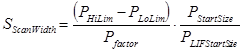
JEOL 8900/8200/8500/8x30 (InterfaceType=2)- The "Peaking Start Size" is used to calculate the spectrometer scan width for the peak scan procedure. The coarse scan width is 3 times larger than the fine scan width.
Where
:
 is the calculated peaking scan width
 is the spectrometer high limit (from MOTORS.DAT)
 is the spectrometer low limit (from MOTORS.DAT)
 is the peak scan size factor (from SCALERS.DAT)
 is the crystal spectrometer peaking size (calculated for 2d and position)
 is the LIF spectrometer peaking size (from SCALERS.DAT)
SX100/SXFive (InterfaceType=5)- The "Peaking Start Size" is modified to produce a number between 0 and 4 by utilizing the following expression :
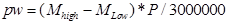
where pw is the SX100/SXFive peak center width
Mhigh is the motor high limit
Mlow is the motor low limit
P is the Peaking Start Size